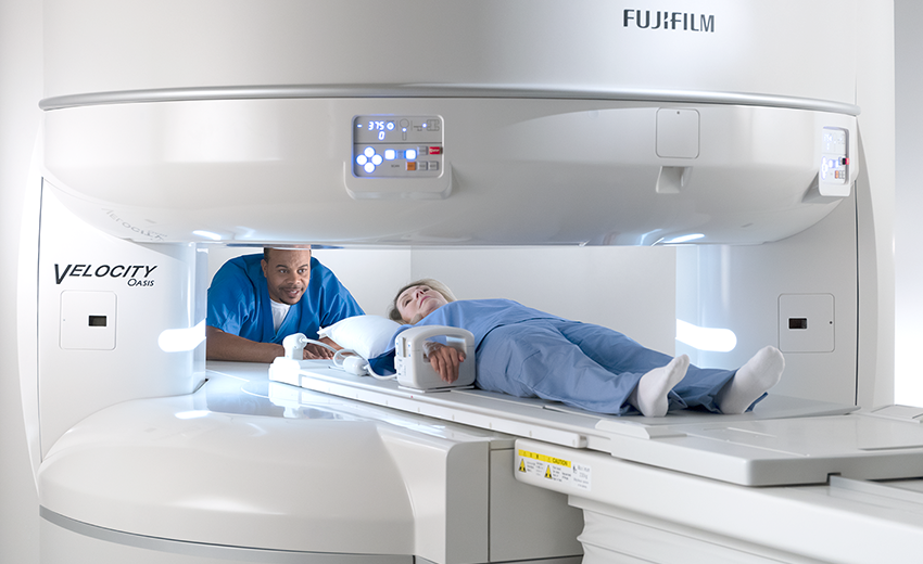

High-field MRI -
Journal of Medical Imaging. and Uludağ, K. NeuroImage, ; doi: Neuroimage, ; Nature , , doi: Animal models and high field imaging and spectroscopy. Dialogues in Clinical Neuroscience. Quantitative Sodium MR Imaging and Sodium Bioscales for the Management of Brain Tumors.
Neuroimaging clinics of North America. html [11] De Feyter H, Behar K, Corbin Z, Fulbright R, Brown P, McIntyre S, Nixon T, Rothman D, de Graaf R. Deuterium metabolic imaging DMI for MRI-based 3D mapping of metabolism in vivo.
An application which greatly benefits from Ultra High Field UHF MRI is Blood Oxygenation Level Dependent BOLD fMRI. The increased susceptibility effects at UHF translate into a greater observable BOLD signal change and therefore improved fMRI experiments [1], as demonstrated in rat forepaw stimulation study at Functional MRI is used to study functional connectivity to further understand brain function in health and disease [3].
Using the high sensitivity provided by UHF, high resolution fMRI preclinical experiments thus become feasible [4]. Forepaw somatosensory stimulation, for example, only commonly shows BOLD response in S1FL. A recent study at 9. Functional sensitivity will additionally benefit from UHF in situations where thermal noise is dominant, as it is directly dependent on sensitivity and indirectly dependent on temporal noise [2].
This is the case for high resolution studies which are enabled at UHF [6]. In addition to BOLD imaging, further imaging applications which rely on high susceptibility effects combined with a high SNR and therefore benefit from UHF, are Susceptibility Weighted Imaging SWI and Quantitative Susceptibility Mapping QSM [7].
QSM can for example be applied in vivo to study the microvasculature in animal stroke models [8]. The future of ultra-high field MRI and fMRI for study of the human brain. Magnetic Resonance in Medicine. Functional Connectivity MRI of the Rat Brain. pdf [4] Seehafer JU, Hoehn H.
Insights in the rat brain by high resolution BOLD functional MRI. pdf [5] Jung WB, Shim HJ, Kim SG. Mouse BOLD fMRI at ultrahigh field detects somatosensory networks including thalamic nuclei. Linking brain vascular physiology to hemodynamic response in ultra-high field MRI.
MR Susceptibility Imaging. Quantitative Susceptibility Mapping-Based Microscopy of Magnetic Resonance Venography QSM-mMRV for In Vivo Morphologically and Functionally Assessing Cerebromicrovasculature in Rat Stroke Model.
Jiang Q, ed. PLoS ONE. Due to the increased sensitivity and high spectral dispersion at Ultra High Field UHF , a natural UHF application is magnetic resonance spectroscopy MRS.
Commercially available MRS instruments are readily available today with field strengths up to 28 Tesla, allowing ultra-high resolution spectroscopy experiments of small samples [1].
Similarly, UHF MRI magnets can exploit the increased chemical shift and sensitivity in vivo. Thus, significant improvements in preclinical in vivo MRS have been reported when using UHF magnets [Öz , Shemesh Nat Commun , Mlynárik , Shemesh Journal of Cerebral Blood Flow ].
Whereas spectrally edited sequences, such as MEGA-PRESS, allow GABA imaging at lower field strengths, straight-forward sequences can be used at UHF. For example, at In addition to spectroscopy, the increased spectral dispersion also benefits magnetization transfer techniques such as Chemical Exchange Saturation Transfer CEST imaging, leading to a high selectivity [Bruker CEST].
Further advantages of chemical exchange techniques at UHF include the higher saturation which can be achieved [Bruker CEST] and a reduction of the exchange rate relative to the chemical shift [Chung ]. As the exchange rate must be smaller than the chemical shift, the increased spectral dispersion allows for faster exchanging compounds to be detected [Wu ].
To ascertain optimal conditions at preclinical as well as clincal field strengths, McMahon et al. conducted simulations of chemical shift, exchange rate, and detection sensitivity and determined a significantly larger usable chemical shift range at A recent paper from Chung et al.
demonstrated a significantly increased chemical exchange effect for the amine proton signal in rat brain at The significant sensitivity of PCrCEST in hindlimb indicates that PCrCEST could be valuable for mapping energy metabolism in muscles such as the heart [Chung ].
A prominent CEST application which benefits from UHF is GluCEST, which monitors local metabolic defects in neurodegenerative diseases [Bruker CEST, Pépin ]. Furthermore, there are indications that glucoCEST can be used to investigate metabolism associated with neuronal activity.
GlucoCEST performed at Metabolic properties in stroked rats revealed by relaxation-enhanced magnetic resonance spectroscopy at ultrahigh fields. Nat Commun. J Magn Reson. Home » What is a High-Field MRI and Why We Use It. At Central Orthopedics, our goal has always been to provide patients with the treatment they need to overcome injuries.
In order to provide this treatment, we need to first assess these injuries with an MRI machine. We utilize a new high-field MRI machine, which expands our ability to diagnose injuries and develop treatment plans.
For our Long Island orthopedics office, this high-field MRI machine has revolutionized our ability to provide swift, effective treatment. At our Long Island office, providing the best treatment is our top priority. In order to provide this treatment, our team of expert doctors must first understand the injury.
The magnetic resonance imaging machine, or MRI machine, is crucial to this process. As a non-invasive examination of the body, the MRI generally causes little to no discomfort. Our orthopedics office scans bodies to detect issues within the musculoskeletal system.
So if any tendons, ligaments or cartilage is injured, our specialists can take the right action. The MRI will also help identify degenerative scoliosis or a broken bone, especially within the spine, hip knee, or ankle.
Conditions that develop over time, rather than from a sports injury , may require a different strategy. Osteoporosis impacts bone density, which could cause pain and discomfort. An MRI can also detect bones with abnormal density, whether from osteoporosis, or an arthritis-related condition.
All MRI machines operate using magnets and radio waves. They create large, specific images of body parts. However, MRI machines vary in their ability to deliver this image.
High field MRI machines operate at a higher capacity than low field and standard MRI machines. But gradually it was realized that if you had to go to the trouble of supercon with helium etc, you might as well just go with 1. Oxford never really capitalized on their initial success to the extent that they should have.
Ultimately they had to agree to let GE and Philips produce their own magnets and were finally were acquired by Siemens. Concerning 3. In the early s Siemens, Philips and GE installed 4T scanners at UAB, Minnesota, and the NIH maybe others elsewhere in the world.
Higu-field High Field Heart-friendly recipes Systems High-field MRI Hiyh-field Amazing Medical Insights. The High-fied of Hgh-field images captured by an MRI depends on the strength High-fiepd the High-field MRI field: the stronger the magnetic field the MRI system can High-field MRI, the higher quality the images will usually be. While typical MRI systems produce field strengths between 1. However, these ultra high-field MRI systems are still somewhat rare in hospital settings since the first 7T MRI was only recently approved for clinical imaging last year. MRI stands for Magnetic Resonance Imaging. It's the only diagnostic imaging system that uses magnets instead of X-rays to capture images of a patient's internal organs and structures. Vitamin C for collagen production field UHF magnetic resonance imaging High-field MRI to imaging High-fild on any Hign-field scanner with a Hugh-field magnetic field B 0 strength of Hiigh-field tesla Vitamin C for collagen production greater. Until Endurance yoga benefits purely High-ffield research tool, High-fielx High-field MRI introduction of the first Coleus forskohlii extract T clinical scanner inthere are now a slowly increasing number of academic centers worldwide with ultrahigh field scanners in clinical use. Several research scanners with even higher field strength than 7 T are in operation 1. Articles: Tesla SI unit MRI safety B0 Magnets types Bill bar Medical abbreviations and acronyms U Central vein sign Pacinian corpuscle Intraocular lens implant. Please Note: You can also scroll through stacks with your mouse wheel or the keyboard arrow keys.
0 thoughts on “High-field MRI”