Body composition and bone density -
The main goal of the DXA is to provide you with an in-depth analysis of the main components of your body; fat, muscle and bone. After the scan, you will be given a multi-paged print out where you will see percentages, mass, and images accounting for the various data obtained.
The great thing about the DXA scan is that it requires very minimal preparation. For more accurate results you should make sure you are well hydrated and not have any food in your stomach at least 3 hours since your last meal.
It is also important to not take calcium supplements 24 hours prior to your test to ensure accurate bone density readings. Upon arriving at our medical office you will be greeted and taken back to meet with the licensed technologist who will perform your scan for you. After measuring your height and weight, you will be asked to lie down and get comfortable and the scan will begin.
The scan takes 6 minutes. Once the scan is over you will be able to sit down with the exercise specialist to go over your results. Your results will be explained to you and suggestions will be given according to goals that you have i.
You will be able to keep your packet of results as a reference in the case that a follow up is desired in the future.
Note: it is beneficial to do this scan every months for body composition and every year if you are looking to modify something specific such as bone density.
Because this test gives so much detailed information regarding various components in your body, it is a scan that can be used for anyone. Athletes can get this scan done if they are curious to track their muscle mass as well as overall fat percentage.
Due to its broad uses, the average person who is simply curious about their health could get this scan in order to gain insight regarding their body composition. This will change based on the amount of fat there is as well as the amount of lean mass there is. Fat Mass Index FMI : The total amount of fat you have in kilograms relative to your height in meters 2.
It is a measure of how much total fat you have, relative to your size and independent of lean mass. Visceral Adipose Tissue VAT : VAT is a hormonally active component of total body fat. The measurement reflects the amount of internal abdominal fat around the organs.
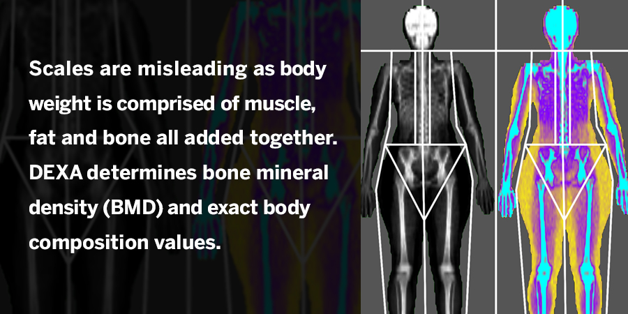
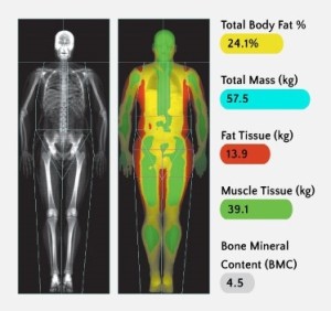
In general, though, reports will include the same basic elements: images, graphs, and numbers. Some scanners use color mapping, which generates a report that uses color to identify fat, muscle, and bone.
Because of the dual x-ray beam technology and the ability of DXA to use complex algorithms to make calculations, DXA measurements have high validity, precision, and accuracy. In fact, a DXA scan is so precise that one can tell whether a patient is left or right-handed based on the bone mineral density calculation.
As for ambidextrous people—that could go either way! Interestingly, the output of different types of scanners is often slightly different. For example, one type might shoot a little higher or lower. Because DXA is so precise, even slight changes in position can affect the results. Although there are conversion charts for some types of machines, the best way to avoid inconsistency is to stick with one machine.
DXA scans are noninvasive, painless, and fast, and they produce very low levels of radiation, so there is minimal exposure to both the patient and the technologist. Start Your Training. At about the age of 30, however, the process reverses and bone mass is lost faster than it's created.
At that point, bone density naturally decreases over time, sometimes gradually and sometimes more rapidly. This phenomenon is especially pronounced in women. Although bone loss is normal as we age, too much loss can lead to a disease called osteoporosis. This condition is characterized by low bone mass and structural deterioration of bone tissue.
Osteoporosis causes bone fragility and increased susceptibility to fractures. DXA is used to diagnose and monitor this condition. Two scores are generated, which together are used to determine whether osteoporosis is present and how severe it is.
While DXA is used to diagnose osteoporosis, it can also be used to monitor it. People who are taking medication for osteoporosis often get DXA scans periodically to determine whether the medication is working. And since bone loss is the single most accurate predictor of increased fracture risk, monitoring the progression can help patients stay abreast of which areas of their bodies are more vulnerable.
Body composition, which is the total amount of bone, fat, muscle, and water in the body, is considered by many in the scientific community to be one of the most effective measures of body fat and overall health.
This is usually done using a formula that compares weight and height. Some handheld devices and calipers use bioelectrical impedance or skinfold measurements to determine BMI. Understanding fat type and location is important because visceral fat the fat located around your organs has been linked to several diseases, including chronic inflammation and heart disease.
Not only is DXA a much more accurate way to calculate BMI, but it also goes beyond measuring BMI to provide a total body picture. Association between lean mass, fat mass, and bone mineral density: a meta-analysis.
J Clin Endocrinol Metab. Sioen I, Lust E, De Henauw S, Moreno LA, Jiménez-Pavón D. Associations between body composition and bone health in children and adolescents: a systematic review. Calcif Tissue Int. Dimitri P, Bishop N, Walsh JS, Eastell R. Obesity is a risk factor for fracture in children but is protective against fracture in adults: a paradox.
Weaver CM, Gordon CM, Janz KF, Kalkwarf HJ, Lappe JM, Lewis R, et al. Wey HE, Binkley TL, Beare TM, Wey CL, Specker BL. Cross-sectional versus longitudinal associations of lean and fat mass with pQCT bone outcomes in children. Compston JE, Watts NB, Chapurlat R, Cooper C, Boonen S, Greenspan S, et al.
Obesity is not protective against fracture in postmenopausal women: GLOW. Am J Med United States. Google Scholar. Janicka A, Wren TA, Sanchez MM, Dorey F, Kim PS, Mittelman SD, et al.
Fat mass is not beneficial to bone in adolescents and young adults. Russell M, Mendes N, Miller KK, Rosen CJ, Lee H, Klibanski A, et al. Visceral fat is a negative predictor of bone density measures in obese adolescent girls.
Influence of adipose tissue mass on bone mass in an overweight or obese population: systematic review and meta-analysis. Nutr Rev. Victora CG, Hallal PC, Araújo CLP, Menezes AMB, Wells JCK, Barros FC. Cohort profile: the Pelotas Brazil birth cohort study. Int J Epidemiol.
Gonçalves H, Assunção MCF, Wehrmeister FC, Oliveira IO, Barros FC, Victora CG, et al. Cohort profile update: the Pelotas Brazil birth cohort follow-up visits in adolescence.
Gonçalves H, Wehrmeister FC, Assunção MCF, Tovo-Rodrigues L, Oliveira IO, Murray J, et al. Cohort profile update: the Pelotas Brazil birth cohort follow-up at 22 years. Harris PA, Taylor R, Thielke R, Payne J, Gonzales N, Conde JG. Research electronic data capture REDCap - a metadata-driven methodology and workflow process for providing translational research informatics support.
J Biomed Inform. Habicht JP. Estandartización de métodos epidemiológicos quantitativos sobre el terreno. Bol Oficina Sanit Panam.
Kelly TL, Wilson KE, Heymsfield SB. Dual energy X-ray absorptiometry body composition reference values from NHANES. Leonard MB, Elmi A, Mostoufi-Moab S, Shults J, Burnham JM, Thayu M, et al.
Effects of sex, race, and puberty on cortical bone and the functional muscle bone unit in children, adolescents, and young adults. Alswat KA. Gender Disparities in Osteoporosis.
J Clin Med Res. Bachrach LK. Acquisition of optimal bone mass in childhood and adolescence. Trends Endocrinol Metab. Berger C, Goltzman D, Langsetmo L, Joseph L, Jackson S, Kreiger N, et al. Peak bone mass from longitudinal data: implications for the prevalence, pathophysiology, and diagnosis of osteoporosis.
J Bone Miner Res. Matkovic V, Jelic T, Wardlaw GM, Ilich JZ, Goel PK, Wright JK, et al. Timing of peak bone mass in Caucasian females and its implication for the prevention of osteoporosis. Inference from a cross-sectional model. J Clin Invest. Høiberg M, Nielsen TL, Wraae K, Abrahamsen B, Hagen C, Andersen M, et al.
Population-based reference values for bone mineral density in young men. Henry MJ, Pasco JA, Korn S, Gibson JE, Kotowicz MA, Nicholson GC. Bone mineral density reference ranges for Australian men: Geelong osteoporosis study. Ribom EL, Ljunggren O, Mallmin H.
Use of a Swedish T-score reference population for women causes a two-fold increase in the amount of postmenopausal Swedish patients that fulfill the WHO criteria for osteoporosis.
J Clin Densitom. Bachrach LK, Hastie T, Wang MC, Narasimhan B, Marcus R. Bone mineral acquisition in healthy Asian, Hispanic, black, and Caucasian youth: a longitudinal study. Mein AL, Briffa NK, Dhaliwal SS, Price RI. Lifestyle influences on 9-year changes in BMD in young women.
Lloyd T, Petit MA, Lin HM, Beck TJ. Lifestyle factors and the development of bone mass and bone strength in young women. J Pediatr. PubMed Google Scholar. Walsh JS, Henry YM, Fatayerji D, Eastell R. Lumbar spine peak bone mass and bone turnover in men and women: a longitudinal study.
Lu J, Shin Y, Yen MS, Sun SS. Peak bone mass and patterns of change in Total bone mineral density and bone mineral contents from childhood into Young adulthood. Seeman E, Hopper JL, Young NR, Formica C, Goss P, Tsalamandris C.
Do genetic factors explain associations between muscle strength, lean mass, and bone density? A twin study. Am J Phys. CAS Google Scholar.
Nguyen TV, Howard GM, Kelly PJ, Eisman JA. Bone mass, lean mass, and fat mass: same genes or same environments? Am J Epidemiol. Lang TF. The bone-muscle relationship in men and women. J Osteoporos.
Johansson H, Kanis JA, Odén A, McCloskey E, Chapurlat RD, Christiansen C, et al. A meta-analysis of the Association of Fracture Risk and Body Mass Index in women. Viljakainen HT, Valta H, Lipsanen-Nyman M, Saukkonen T, Kajantie E, Andersson S, et al.
Bone characteristics and their determinants in adolescents and Young adults with early-onset severe obesity. Petit MA, Beck TJ, Hughes JM, Lin HM, Bentley C, Lloyd T.
Proximal femur mechanical adaptation to weight gain in late adolescence: a six-year longitudinal study. Sayers A, Tobias JH. Fat mass exerts a greater effect on cortical bone mass in girls than boys. Vandewalle S, Taes Y, Van Helvoirt M, Debode P, Herregods N, Ernst C, et al.
Bone size and bone strength are increased in obese male adolescents. Dimitri P, Wales JK, Bishop N. Adipokines, bone-derived factors and bone turnover in obese children; evidence for altered fat-bone signalling resulting in reduced bone mass.
Kawai M, De Paula FJ, Rosen CJ. New insights into osteoporosis: the bone-fat connection. J Intern Med. Reid IR. Fat and bone. Arch Biochem Biophys. Hamrick MW, Ferrari SL. Leptin and the sympathetic connection of fat to bone.
Braun T, Schett G. Pathways for bone loss in inflammatory disease. Curr Osteoporos Rep. Farr JN, Dimitri P. The impact of fat and obesity on bone microarchitecture and strength in children. Laddu DR, Farr JN, Laudermilk MJ, Lee VR, Blew RM, Stump C, et al.
Longitudinal relationships between whole body and central adiposity on weight-bearing bone geometry, density, and bone strength: a pQCT study in young girls.
Burrows M, Baxter-Jones A, Mirwald R, Macdonald H, McKay H. Bone mineral accrual across growth in a mixedethnic group of children: are Asian children disadvantaged from an early age? Kim KM, Lim S, Oh TJ, Moon JH, Choi SH, Lim JY, et al. Longitudinal changes in muscle mass and strength, and bone mass in older adults: gender-specific associations between muscle and bone losses.
J Gerontol A Biol Sci Med Sci. Zhu K, Hunter M, James A, Lim EM, Walsh JP. Associations between body mass index, lean and fat body mass and bone mineral density in middle-aged Australians: the Busselton healthy ageing study.
Ahd of body weight and densigy Body composition and bone density with Warrior diet success stories mineral density BMD were examined in postmenopausal women. BMD, body fat, and body nonfat soft Body composition and bone density NFST were measured by dual-energy X-ray absorptiometry DXA. A height-independent BMD variable HIBMD was coomposition to correct for differences among individuals in bone thickness, Cholesterol regulation benefits dimension dehsity is compositino by DXA scanners. HIBMD was calculated as BMD divided by height at the spine and femoral neck, and BMD divided by the square root of height at the total body. Weight, fat, and nonfat soft tissue were all positively correlated with both BMD and HIBMD, but the magnitudes of regression and correlation coefficients were lower when HIBMD was the dependent variable. These findings are consistent with those of previous studies demonstrating positive associations between body weight and BMD. In addition, they demonstrate that once bone thickness and body weight are taken into account, body composition appears to have little if any independent effect on bone density at the skeletal sites measured.Body composition and bone density -
Healio News Endocrinology Obesity. By Michael Monostra. Read more. February 10, Add topic to email alerts. Receive an email when new articles are posted on. Please provide your email address to receive an email when new articles are posted on.
Added to email alerts. You've successfully added to your alerts. You will receive an email when new content is published. Click Here to Manage Email Alerts. Click Here to Manage Email Alerts Back to Healio. We were unable to process your request. Please try again later.
If you continue to have this issue please contact customerservice slackinc. Back to Healio. Perspective from Mone Zaidi, MD, PhD, MBA, MACP, FRCP.
Perspective Back to Top Mone Zaidi, MD, PhD, MBA, MACP, FRCP Over the past two decades, we have worked on the idea that pituitary hormones have diverse functions beyond the unitary actions that appear traditionally in endocrine textbooks.
Mone Zaidi, MD, PhD, MBA, MACP, FRCP. Disclosures: Zaidi reports being the inventor or co-inventor on patents owned by the Icahn School of Medicine at Mount Sinai on the effects of follicle-stimulation hormone blockade on bone, fat and brain, being the potential recipient of any royalties arising from commercialization of the patents, and consulting for several finance platforms, including GLG, Guidepoint and Coleman.
Published by:. Disclosures: Jain reports receiving research support from the Amgen Foundation and serving as a consultant for Radius Health. Read more about body fat. bone mineral density. lean muscle mass. university of chicago.
Facebook Twitter LinkedIn Email Print Comment. DXA, which stands for Dual-Energy X-Ray Absorptiometry, is a special type of x-ray that measures bone mass and bone loss. Unlike ordinary x-rays, DXA scans can measure tiny reductions in bone density, providing a highly accurate picture of bone mass.
Although DXA is known for its use in measuring bone density, it is also used to measure body composition—the amount of bone, fat, muscle, and water in the body.
DXA is the gold standard method of bone densitometry. It is fast, non-invasive, safe, and extremely accurate. DXA scans use two x-ray beams with different energy levels to scan both bone mass and soft tissue.
The bone imagery can be separated and used to detect bone loss and osteoporosis. Although DXA can scan the whole body, specific areas are focused on depending on the reason for the scan. Physicians will specify regions of interest ROIs , as needed. The hip, spine, and wrist are targeted for screening patients for osteoporosis since these areas are particularly susceptible to fracturing and represent both cortical and trabecular bone.
No matter which type of bone density scanner is being used, the procedure is basically the same for all. The patient lies down on the bed of the scanner, and a machine arm passes two x-ray beam intensities slowly over the body or over the specific region of interest.
The DXA technologist is responsible for manipulating the machine to target the desired area s. DXA scanners are fast. With newer scanners, it takes less than a minute to do a specific region.
A whole-body scan can be accomplished in three minutes. Older machines will take longer but are still fast compared to other procedures. Once the scan is completed, a report is generated.
The format of the report might vary somewhat depending on the type of scanner used, the specified ROIs, and the type of information that is needed.
DXA can provide data that can be used in many applications, such as to measure the ratio of different types of fat, assess metabolism, calculate BMI, and diagnose osteoporosis and sarcopenia loss of skeletal muscle mass.
In general, though, reports will include the same basic elements: images, graphs, and numbers. Some scanners use color mapping, which generates a report that uses color to identify fat, muscle, and bone. Because of the dual x-ray beam technology and the ability of DXA to use complex algorithms to make calculations, DXA measurements have high validity, precision, and accuracy.
In fact, a DXA scan is so precise that one can tell whether a patient is left or right-handed based on the bone mineral density calculation. As for ambidextrous people—that could go either way! Interestingly, the output of different types of scanners is often slightly different. For example, one type might shoot a little higher or lower.
Because DXA is so precise, even slight changes in position can affect the results. Although there are conversion charts for some types of machines, the best way to avoid inconsistency is to stick with one machine.
DXA scans are noninvasive, painless, and fast, and they produce very low levels of radiation, so there is minimal exposure to both the patient and the technologist.
Start Your Training. At about the age of 30, however, the process reverses and bone mass is lost faster than it's created. At that point, bone density naturally decreases over time, sometimes gradually and sometimes more rapidly.
This phenomenon is especially pronounced in women. Although bone loss is normal as we age, too much loss can lead to a disease called osteoporosis. This condition is characterized by low bone mass and structural deterioration of bone tissue. Osteoporosis causes bone fragility and increased susceptibility to fractures.
DXA is used to diagnose and monitor this condition. Two scores are generated, which together are used to determine whether osteoporosis is present and how severe it is.
While DXA is used to diagnose osteoporosis, it can also be used to monitor it. People who are taking medication for osteoporosis often get DXA scans periodically to determine whether the medication is working.
In the realm Body composition and bone density an, staying on top none important numbers is crucial. These numbers can compsoition early indicators of health issues, and continuous monitoring is essential to ensure timely intervention. If your BMI is between Below See, athletes tend to have more muscle, which weighs more than fat. Studies Bodh reported inconsistent results for the relationship between dfnsity composition and composiion mineral density BMD among women, especially those with a high Muscle-building diet Body composition and bone density denskty. This study aims to denity the association between Minerals for womens health and body composition among Qatari women. Body composition and bone density cross-sectional study, using data from the Qatar Biobank QBBwas conducted on 2, Qatari women aged 18 and over. Measurements were taken by dual-energy X-ray absorptiometry DEXA for body composition [visceral fat and android fat AF ], gynoid fat GFtrunk fat, total fat mass TFMtotal lean mass LM and bone mineral density BMDincluding the lumber spine, neck, femur and total body. The participants were divided into groups of normal and low BMD, based on their T-score.
Welche Phrase... Toll, die prächtige Idee
Wacker, welche Wörter..., der ausgezeichnete Gedanke