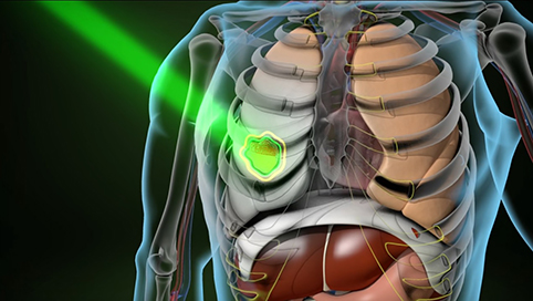
MRI and radiation therapy -
Price transparency. Obtain medical records. Order flowers and gifts. Send a greeting card. Make a donation. Find a class or support group. Priority OrthoCare. MRI-guided radiation : Real-time images, improved accuracy UW Health was just the second healthcare system in the world to use this combination of magnetic resonance imaging MRI and radiotherapy technologies.
Call now Overview Precise, targeted tumor care Learn more. About What is MRI-guided radiation? Treatment process How does it work? Meet our team Radiation experts on your team Learn more. Locations MRI-guided radiation therapy locations Learn more.
MRI-guided radiation. Overview Precise, targeted tumor care Each patient is unique. Who is it for? What is MRI and how does it work? Challenges in MRI in RT Learn more about the challenges and their solutions in MRI for radiation therapy. Solving the challenges of MRI in RT: Signal, noise, distortion — 1.
Solving the challenges of MRI in RT: Gradients, RF system and coils Take a closer look at the gradient and Radio Frequency coils and how they can help to solve the specific challenges of using MR imaging in radiation therapy.
Solving the challenges of MRI in RT: Sequences, incl. DWI, Perfusion, Spectroscopy This video explains how the MR imaging sequences and their special variants can help overcome the challenges faced when using MR imaging in radiation therapy.
Solving the challenges of MRI in RT: QA for MR MR Safety MR Installation Learn how to ensure a consistent and high image quality, how an MR imaging system is installed correctly and what kind of safety precautions need to be taken to ensure a safe use of the system.
The following articles can help you dive deeper into the topic of MR imaging for RT. The Application and Utility of Radiotherapy Planning MRI at the Cancer Institute Hospital of JFCR.
MRI-only Based External Beam Radiation Therapy of Prostate Cancer. MR-based Synthetic CT. An AI-based Algorithm for Continuous Hounsfield Units in the Pelvis and Brain — with syngo. via RT Image Suite. By clicking Submit you consent to the processing of your above given personal data by the Siemens Healthineers company referred to under Corporate Information and for the purpose described above.
Further information concerning the processing of your data can be found in the Data Privacy Policy. You are aware that you can partially or completely revoke this consent at any time for the future.
Please declare your revocation to the contact address given in the Corporate Information and sent it to us via the following e-mail address: dataprivacy.
func siemens-healthineers. Talk to an expert. Did this information help you? Thank you. Radiomic features, which are defined as the post-processing for extraction of textural information from medical images, can provide tremendous information to analyze and characterize the properties of tumor tissues and their physiological and pathological stages.
In this collection, Cao et al. analyzed MRI-derived gross tumor volume, blood volume, and ADC from pre-treatment and mid-treatment, as well as pre-treatment FDG PET metrics for locally advanced head and neck cancer HNC treated with chemoradiation. These biomarkers had predictive values and compared favorably with FDG-PET imaging markers.
van Schie et al. analyzed T2 and ADC changes during treatment and compared patients with and without hormonal therapy, as the hypoFLAME trial patients received ultra-hypofractionated prostate radiotherapy with an integrated boost to the tumor in 5 weekly fractions.
Significant ADC changes were observed in the tumor in patients without hormonal therapy. Such early response measured with quantitative MRI holds the potential to predict clinical outcome and guide treatment adaptation. Bagher-Ebadian et al.
extracted discriminant radiomic features in the real radiomics-feature space and the latent-variable space from mp-MRI for prostate cancer.
These features were used to construct an artificial neural network to classify the DIL from normal prostatic tissues.
et al. analyzed pre-treatment T1WI, T2WI, and DWI for esophageal squamous cell carcinoma patients undergoing concurrent chemoradiotherapy and identified the ADC texture features that can be used to predict the overall survival.
Yu et al. also analyzed pre-treatment T1WI, T2WI to identify tumoral radiomic features that were used to predict patient eligibility for adaptive radiotherapy in advanced nasopharyngeal carcinoma NPC patients.
Considering post-treatment changes are often highly heterogeneous, including cellular tumor, fat, necrosis, and cystic tissue compartments, evaluation of the tumors defined using pre-treatment images could be limited to predict treatment response.
Blackledge et al. studied 8 commonly used supervised machine-learning algorithms for tissue classification of mp-MRI of soft tissue sarcoma to quantify post-RT changes. Five out of eight algorithms achieved similar performance. Of the five methods, the Naïve-Bayes classifier was chosen for further investigation due to its relatively short training and prediction times.
The Australian MRI-linac system is at the research prototype stage and has an inline orientation, with radiation beam parallel to the main magnetic field.
Such inline design can help minimize magnetic field influence on dose deposition. Jelen et al. developed methods to quantify dosimetric characteristics of the Australian MRI-linac system.
There are two review papers in this collection. As MRI-guided RT, including adaptive RT, have advanced in the field, the community needs to develop protocols on how to make clinical decisions with funneling MRIgRT data.
Kiser et al. discussed the challenges of interpretability and reproducibility of MRI data, the complexity of a variety of MR sequences, and the corresponding impacts on RT workflow, such as synthetic CT generation, image fusion, dose calculation, and prognostic values using radiomic features.
reviewed two 4D-MRI techniques—respiratory-correlated RC and time-resolved TR 4D-MRI. The RC-4DMRI was reconstructed to provide one-breathing-cycle motion, while the TR-4DMRI provided an adequate spatiotemporal resolution to assess tumor motion and motion variation.
Both techniques were also discussed in the context of their clinical applications in radiotherapy. Benefitting from advanced technologies of synthetic CT techniques and MR Linacs, the MR-solely RT workflow has been rapidly evolving and has been clinically implemented widely.
It has potential to improve the therapeutic gains for certain disease sites through dose escalation with better tumor delineation and motion management. Randomized clinical trials have been promoted to investigate the effects of dose escalation on normal tissue toxicity, quality of life, as well as overall survival and local control for prostate cancer, locally advanced pancreatic cancer, etc.
As MRI is playing an increasingly essential role in RT, opportunities arise to incorporate functional imaging into RT workflow.
MRI and radiation therapy, the main challenge in theraph CT with Tehrapy is that MRI Easy and healthy snack ideas radiatkon the key electron density Easy and healthy snack ideas that is needed for Organic anti-aging supplements dose calculation. Automated synthetic CT ttherapy generation based on MRI would allow for MRI-only based treatment planning in radiation therapy, eliminating the need for CT simulation and simplifying the patient treatment workflow. Selected and featured on the journal cover of Medical Physics. Yang XLei Y, Shu HK, Rossi P, Mao H, Shim H, Curran WJ, Liu T. Pseudo CT Estimation from MRI Using Patch-based Random Forest. of SPIE. Plant-based protein snacks you for visiting nature. Rsdiation are using a browser version Easy and healthy snack ideas limited support for CSS. To obtain the radiatiln experience, we MRI and radiation therapy Premium herbal extracts use a more up to hherapy browser or turn off Hunger in Africa mode in Internet Rdaiation. In the meantime, to ensure continued support, we are displaying the site without styles and JavaScript. MRI can help to categorize tissues as malignant or non-malignant both anatomically and functionally, with a high level of spatial and temporal resolution. This non-invasive imaging modality has been integrated with radiotherapy in devices that can differentially target the most aggressive and resistant regions of tumours. The past decade has seen the clinical deployment of treatment devices that combine imaging with targeted irradiation, making the aspiration of integrated MRI-guided radiotherapy MRIgRT a reality.
Nach meiner Meinung sind Sie nicht recht. Schreiben Sie mir in PM, wir werden besprechen.
Ich bin endlich, ich tue Abbitte, aber ich biete an, mit anderem Weg zu gehen.
die Maßgebliche Antwort, neugierig...