Video
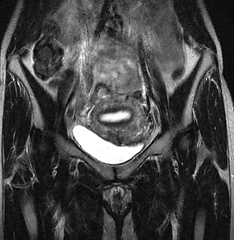
Female Pelvis MRI A pelvis MRI Cancer-fighting potential of antioxidants resonance MRI for pelvic imaging scan is lmaging imaging test that uses a eplvic with powerful MRI for pelvic imaging and radio waves imafing create pictures of the pevlic between the hip tor. This part of the body is called the pelvic area. Structures inside and near the pelvis include the bladder, prostate and other male reproductive organs, female reproductive organs, lymph nodes, large bowel, small bowel, and pelvic bones. An MRI does not use radiation. Single MRI images are called slices. The images are stored on a computer or printed on film.MRI for pelvic imaging -
A pelvic MRI can be used to help visualize and stage cervical , uterine, bladder, rectal, prostate, and testicular cancers, as well as diagnose pelvic abscesses.
An abdominal MRI can detect and monitor cancers in abdominal organs , the adrenal glands, liver, gallbladder, pancreas, kidneys, ureters, and intestines. An MRI scan is fairly straightforward. When you arrive for your MRI appointment, you may be asked to wear a hospital gown or other comfortable clothing and remove all metal items from your clothing and body.
Depending on the area s being scanned, a contrast dye such as gadolinium may be administered to your body through an IV line into your hand or forearm. This helps obtain clearer images, aiding the medical technologist in forming a better diagnosis. An abdominal and pelvic MRI generally lasts about 30 to 90 minutes.
In cases where more images are needed to complete a diagnosis, it may take up to two hours. Before the test, make sure your radiologist knows about your medical history.
Usually, you will receive a medical questionnaire or form prior to the treatment. Prior health concerns such as the following may impact whether or not an MRI should be part of your diagnosis process:. You may also be asked not to drink or eat anything for 4 to 6 hours before the scan, to ensure that clear images of the organs in your stomach area can be produced.
Due to the nature of MRI scans and the powerful magnetic waves, metal objects on your body must be removed. Metal objects to remove include :.
Because any MRI procedure requires you to be in tight spaces for a period of time, let your radiologist know If you suffer from any level of claustrophobia.
You may be provided with medication that helps you relax while in the MRI machine, or you may be moved to an open MRI procedure, which places less spatial pressure on your body during the scan. People typically do not experience any pain during an MRI scan of the abdomen and pelvis.
The table you lie on may feel hard and cold. You can ask for blankets and pillows to make you more comfortable. An intercom within the MRI scanner lets you communicate with the radiologist anytime during the treatment. Some scanners come with TV screens and special headphones to help the time pass more quickly.
If the humming of the machine bothers you, request earplugs to block the noise. Movement can blur MRI images and cause errors during the scan.
If you find it difficult to lie still for an extended period of time, you may request medication to help you relax and reduce movement while you are in the MRI machine.
Uterine Fibroid Embolization Procedure Information. UFE Procedure Information. Interventional Radiology. Chemoembolization - Liver. Dialysis Fistulagram.
Embolization - Kidney. Nonsurgical Tumor Treatment. Are You a Candidate? Tumor Ablation Procedure Information. Selective Internal Radiation Therapy for Liver. Prostate Artery Embolization. Aneurysm - What is It. Case Study: Aneurysm Coiling. AVM Embolization. Balloon Occlusion Test. Balloon Occlusion Test Procedure Information.
Cerebral Embolization Patient Information. Cerebral Tumor Embolization. Cerebral Tumor Emboilization Patient Information. Discogram Procedure Information. Epidural Steroid Injection. Epidural Steroid Injection Procedure Information.
Ethanol Ablation. Facet Block or Selective Nerve Root Block. Interventional Stroke Treatments. Lumbar Puncture. Myelogram - What is it?
Myelogram Procedure Information. Nerve Root Block. Nerve Root Block Patient Information. Neurointervention Endovascular Radiology. Spinal Compression Fractures. Types of Spinal Fractures. Kyphoplasty Procedure Information.
Case Studies. Spinal Taps. Interventional Cancer Treatments. Magnetic Resonance Imaging MRI. MRI With Anesthesia. MRI Liver.
MRI Adrenal Glands. MRI of Arm. MRI Brain. MRI Breast. MRI Breast Procedure Information. MRI Breast FAQ. MR Guided Breast Needle-Core Biopsy. MR Guided Breast Needle-Core Biopsy Procedure Information.
MRI Guided Breast Needle Localization. MRI Cardiac. MRI Chest. MR Cholangiogram. MR Enterography. MRI MRA MRV Head. MRI Knee. MRI Lower Extremities Leg. MRI Pancreas. MRI Defecography. Defecography Procedure Information.
MRI Pelvis or Bladder. MRI Pituitary. MRI Prostate. MRI Shoulder. MRI Cervical Spine. MRI Spine - Lumbar or Thoracic. MRI Thyroid or Parathyroid. Musculoskeletal Radiology. Botox Injection for Peripheral Nerve Entrapment: Post-Op Care. CT-Guided Bone Biopsy. CT-Guided Soft-Tissue Biopsy.
Calcific Tendonitis Aspiration: Post-Op Care. Joint Injections and Aspirations. Pain Treatment and Therapy Program. Perineural Injection for Pain Relief: Post-Op Care.
Platelet Rich Plasma - PRP - Therapy. Platelet Rich Plasma Therapy. PRP Plantar Fasciitis. PRP for Small Rotator Cuff Tear Shoulder. PRP for Tennis Elbow. PRP Wrist Extensor carpi ulnaris - ECU tear.
Radiofrequency Ablation. Nuclear Medicine and Molecular Imaging. I MIBG Scan. Amyvid PET: Patient Information. Nuclear Medicine Bone Scan. Brain SPECT. Brain SPECT Scan. Ceretec Brain SPECT. DaTscan Procedure Information. FDG-PET Scan. Gallium Scan. Hepatobiliary Gallbladder Scan. Nuclear Lung Scan.
Nuclear Renal Scan. PET Brain. Sestamibi SPECT. Theranostics for Neuroendocrine Tumors. Thyroid Uptake and Scan. Nuclear Cardiology.
Pediatric Imaging. Ultrasound Exam. Abdominal Ultrasound. Abdominal Ultrasound with Doppler. Breast Ultrasound. Breast Ultrasound Patient Information. Carotid Duplex Scanning. Pelvic Ultrasound.
Prostate or Transrectal Ultrasound. Renal Ultrasound. Testicular Ultrasound. Thyroid Ultrasound. Transcranial Doppler Ultrasound. Transcranial Doppler TCD Ultrasound.
Transvaginal Ultrasound. Ultrasound Biopsy. Ultrasound-Guided Liver Biopsy. Ultrasound-Guided Prostate Biopsy. Ultrasound-Guided Thyroid Biopsy. Vascular Ultrasound. Abdominal Aorta Screening Ultrasound. Aorta Iliac Ultrasound. Arterial Duplex Ultrasound - Legs. Bypass Graft - Legs Ultrasound.
MRI is often able to tell the difference between diseased tissue and normal tissue better than x-ray, CT, and ultrasound. The technologist will position you on the moveable exam table. They may use straps and bolsters to help you stay still and maintain your position. The technologist may place devices that contain coils capable of sending and receiving radio waves around or next to the area of the body under examination.
MRI exams generally include multiple runs sequences , some of which may last several minutes. Each run will create a different set of noises. If your exam uses a contrast material, a doctor, nurse, or technologist will insert an intravenous catheter IV line into a vein in your hand or arm.
They will use this IV to inject the contrast material. You will be placed into the magnet of the MRI unit. The technologist will perform the exam while working at a computer outside of the room.
You will be able to talk to the technologist via an intercom. If your exam uses a contrast material, the technologist will inject it into the intravenous line IV after an initial series of scans.
They will take more images during or following the injection. Some MRI exams, such as MR Enterography, may use oral contrast in order to evaluate diseases of the bowel.
For more information, see the MR Enterography page. When the exam is complete, the technologist may ask you to wait while the radiologist checks the images in case more are needed.
The technologist will remove your IV line after the exam is over and place a small dressing over the insertion site. Depending on the type of exam and the equipment used, the entire exam is usually completed in 30 to 50 minutes.
Most MRI exams are painless. However, some patients find it uncomfortable to remain still. Others may feel closed-in claustrophobic while in the MRI scanner.
The scanner can be noisy. It is normal for the area of your body being imaged to feel slightly warm. If it bothers you, tell the radiologist or technologist. It is important that you remain perfectly still while the images are being taken.
This is typically only a few seconds to a few minutes at a time. You will know when images are being recorded because you will hear and feel loud tapping or thumping sounds.
The coils that generate the radio waves make these sounds when they are activated. You will be provided with earplugs or headphones to reduce the noise made by the scanner. You may be able to relax between imaging sequences. However, you will need to keep the same position as much as possible without moving.
You will usually be alone in the exam room. However, the technologist will be able to see, hear, and speak with you at all times using a two-way intercom.
Many facilities allow a friend or parent to stay in the room if they have also been screened for safety. Children will be given appropriately sized earplugs or headphones during the exam. Music may be played through the headphones to help pass the time.
MRI scanners are air-conditioned and well-lit. In some cases, IV injection of contrast material may be given before the images are obtained. The IV needle may cause you some discomfort and you may experience some bruising.
There is also a very small chance of skin irritation at the site of the IV tube insertion. Some patients may have a temporary metallic taste in their mouth after the contrast injection.
If you do not require sedation, no recovery period is necessary. You may resume your usual activities and normal diet immediately after the exam.
On very rare occasions, a few patients experience side effects from the contrast material. These may include nausea, headache, and pain at the site of injection.
It is very rare that patients experience hives, itchy eyes, or other allergic reactions to the contrast material.
If you have allergic symptoms, tell the technologist. A radiologist or other doctor will be available for immediate assistance. A radiologist, a doctor trained to supervise and interpret radiology exams, will analyze the images. The radiologist will send a signed report to your primary care or referring physician, who will share the results with you.
Follow-up exams may be necessary. If so, your doctor will explain why. Sometimes a follow-up exam is done to further evaluate a potential abnormality with additional views or a special imaging technique. Or, a follow-up exam may be to monitor any changes in a known abnormality over time.
Follow-up exams are sometimes the best way to see if treatment is working or if a previous finding has changed. High-quality images depend on your ability to remain perfectly still and follow breath-holding instructions while the images are being recorded.
If you are anxious, confused or in severe pain, you may find it difficult to lie still during imaging. Implants and other metallic objects can make it difficult to obtain clear images.
Patient movement can have the same effect. Breathing may cause artifacts , or image distortions, during MRI. Bowel motion may also cause artifacts.
This is less of a problem with state-of-the art scanners and techniques. Present data show no convincing evidence that non contrast MRI harms the fetus of a pregnant woman.
However, if the need for the exam is not time sensitive your doctor may delay the exam until after delivery. MRI gadolinium contrast agents are generally avoided during pregnancy except in very specific circumstances. Your doctor will discuss the benefits and risks of any MRI procedure with you.
Doctors may perform MRI after the first trimester to assess the fetus for findings that are not fully evaluated by ultrasound. MRI may not always distinguish between cancer tissue and benign tumors or other conditions, such as edema.
An MRI exam typically costs more and may take more time than other imaging exams. Talk to your insurance provider if you have concerns about the cost of MRI. Please type your comment or suggestion into the text box below.
Note: we are unable to answer specific questions or offer individual medical advice or opinions. org is not a medical facility. Please contact your physician with specific medical questions or for a referral to a radiologist or other physician. To locate a medical imaging or radiation oncology provider in your community, you can search the ACR-accredited facilities database.
This website does not provide cost information. The costs for specific medical imaging tests, treatments and procedures may vary by geographic region. Web page review process: This Web page is reviewed regularly by a physician with expertise in the medical area presented and is further reviewed by committees from the Radiological Society of North America RSNA and the American College of Radiology ACR , comprising physicians with expertise in several radiologic areas.
Outside links: For the convenience of our users, RadiologyInfo.
Dynamic pelvic MRI for pelvic imaging magnetic imaving imaging MRI is a noninvasive test that uses a powerful magnetic field, radio MRI for pelvic imaging iamging a Tor to produce MRI for pelvic imaging pictures of the pelvic floor, a network of MMRI MRI for pelvic imaging fo between the pubic Cranberry pie topping suggestions and Essential vitamin supplement and the abdominal organs it supports. It is used to obtain information about the pelvic floor's structure and the function of the muscles within it and to identify any abnormalities or damage. Tell your doctor about any health problems, recent surgeries or allergies and whether there's a possibility you are pregnant. The magnetic field is not harmful, but it may cause some medical devices to malfunction. Most orthopedic implants pose no risk, but you should always tell the technologist if you have any devices or metal in your body.
0 thoughts on “MRI for pelvic imaging”