
Video
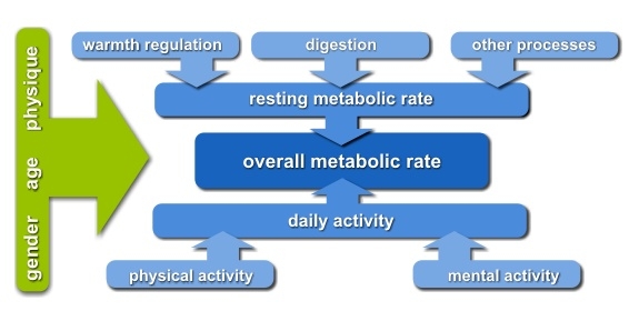
Resting Metabolic RateMetabolic rate regulation -
Some of the more common hormonal disorders affect the thyroid. This gland secretes hormones to regulate many metabolic processes, including energy expenditure the rate at which kilojoules are burned.
Thyroid disorders include:. Our genes are the blueprints for the proteins in our body, and our proteins are responsible for the digestion and metabolism of our food. Sometimes, a faulty gene means we produce a protein that is ineffective in dealing with our food, resulting in a metabolic disorder.
In most cases, genetic metabolic disorders can be managed under medical supervision, with close attention to diet.
The symptoms of genetic metabolic disorders can be very similar to those of other disorders and diseases, making it difficult to pinpoint the exact cause.
See your doctor if you suspect you have a metabolic disorder. Some genetic disorders of metabolism include:. This page has been produced in consultation with and approved by:. Content on this website is provided for information purposes only.
Information about a therapy, service, product or treatment does not in any way endorse or support such therapy, service, product or treatment and is not intended to replace advice from your doctor or other registered health professional.
The information and materials contained on this website are not intended to constitute a comprehensive guide concerning all aspects of the therapy, product or treatment described on the website.
All users are urged to always seek advice from a registered health care professional for diagnosis and answers to their medical questions and to ascertain whether the particular therapy, service, product or treatment described on the website is suitable in their circumstances.
The State of Victoria and the Department of Health shall not bear any liability for reliance by any user on the materials contained on this website. Skip to main content. Actions for this page Listen Print. Summary Read the full fact sheet. On this page. What is metabolism?
Two processes of metabolism Metabolic rate Metabolism and age-related weight gain Hormonal disorders of metabolism Genetic disorders of metabolism Where to get help.
Two processes of metabolism Our metabolism is complex — put simply it has 2 parts, which are carefully regulated by the body to make sure they remain in balance. They are: Catabolism — the breakdown of food components such as carbohydrates , proteins and dietary fats into their simpler forms, which can then be used to provide energy and the basic building blocks needed for growth and repair.
Anabolism — the part of metabolism in which our body is built or repaired. Anabolism requires energy that ultimately comes from our food.
When we eat more than we need for daily anabolism, the excess nutrients are typically stored in our body as fat. Thermic effect of food also known as thermogenesis — your body uses energy to digest the foods and drinks you consume and also absorbs, transports and stores their nutrients.
Energy used during physical activity — this is the energy used by physical movement and it varies the most depending on how much energy you use each day. Physical activity includes planned exercise like going for a run or playing sport but also includes all incidental activity such as hanging out the washing, playing with the dog or even fidgeting!
Basal metabolic rate BMR The BMR refers to the amount of energy your body needs to maintain homeostasis. Factors that affect our BMR Your BMR is influenced by multiple factors working in combination, including: Body size — larger adult bodies have more metabolising tissue and a larger BMR.
Amount of lean muscle tissue — muscle burns kilojoules rapidly. Crash dieting, starving or fasting — eating too few kilojoules encourages the body to slow the metabolism to conserve energy.
Age — metabolism slows with age due to loss of muscle tissue, but also due to hormonal and neurological changes. Growth — infants and children have higher energy demands per unit of body weight due to the energy demands of growth and the extra energy needed to maintain their body temperature.
Gender — generally, men have faster metabolisms because they tend to be larger. Genetic predisposition — your metabolic rate may be partly decided by your genes. Hormonal and nervous controls — BMR is controlled by the nervous and hormonal systems.
Thus, we included a total of 21 papers in our analyses, of which 12 were on birds, and 9 on mammals. Nine of the 22 papers included more than one experimental treatment, yielding a total of 35 effect sizes.
For each of these studies, we extracted information on study species or metabolic and GC variables reported, among others Table S2. all individuals went through all experimental treatments ; c Whether metabolic rate or heart rate was the metabolic variable; and d The type of treatment that induced an increase in metabolic rate see below Table S2.
To estimate effect sizes of metabolism and GCs, we used the web-based effect size calculator Practical Meta-Analysis Effect Size Calculator www. See Table S3 for details on data extraction and effect size calculations. For each study, we compared the mean metabolic rate and level of plasma GCs of individuals in the treatment group s to that of individuals in the control group.
For studies in which treatment was confounded with time, because pre-treatment measurements were used as control and compared with measurements during treatment, the pre-treatment measure was used as control when calculating effect sizes in studies where there was a single treatment.
When studies with a before-after design included more than one experimental treatment, the treatment yielding the lowest metabolism was taken as control for the effect size calculations. Thus, confounding time with treatment was avoided whenever possible.
We conducte d all meta-analyses using the rma. mv function from the metafor package Viechtbauer , implemented in R version 4. Standard errors were used for the weigh factor.
All models contained a random intercept for study identity to account for inclusion of multiple experimental treatments or groups from the same study. Most species were used in a single study, and we therefore did not include species as a random effect in addition to study identity. The number of species was however insufficient to reliably estimate phylogenetic effects, we therefore limited the analysis in this respect with a comparison between birds and mammals see below.
The dependent variable was either the metabolic rate or the GC effect size. One model was fitted with the metabolic rate effect size as a dependent variable, to estimate the average effect on metabolic rate across all studies in the analyses.
All other models had the GC effect size as dependent variable, and metabolic rate effect size as a moderator. Distribution of metabolic rate effect sizes was skewed which was resolved by ln-transformation, which yielded a better fit when compared to a model using the linear term evaluated using AIC, see results for details.
Our first GC model contained only the metabolic rate effect size as a fixed independent variable. This model provides a qualitative test of whether GC levels increase when metabolic rate is increased and tests prediction i by providing an estimate of the intercept, which represents the average GC effect size because we mean centered the ln-transformed metabolic rate effect size Schielzeth The same model tests prediction ii whether the GC effect increases with an increasing metabolic rate effect size, which will be expressed in a significant regression coefficient of the metabolic rate effect size.
Following the model with which we tested our main predictions, we ran additional models to test for the effects on GC effect sizes of a taxa birds vs. This last factor tests our prediction iii.
We included these variables as modulators in the analysis, as well as the two-way interactions of these factors with the metabolic rate effect size.
Treatment type was categorized as 1 climate , 2 psychological , or 3 others. We compared models with vs. To rule out publication bias effects i.
Variable effects and results remained quantitatively very similar and qualitatively unchanged. However, this variable had a negligible effect on the models, and we therefore excluded it from the final models. Among the studies selected for inclusion in the analysis, the treatment effect size on metabolic rate MR was on average 1.
There was a strong association between MR effect sizes and GC effect sizes Table 1 , Fig. It is further worth noting that the residual heterogeneity did not exceed the level expected by chance Table 1.
Meta regression model testing the association between metabolic rate MR effect sizes and glucocorticoid effect sizes. Area of dots is proportional to the experiment sample size i.
square root of the number of individuals in which GCs were measured. Furthermore, none of these variables had a significant effect on GC effect size, nor did the association between MR and GC effect sizes depend on those factors i.
interactions between these variables and MR effect sizes were always non-significant; Table 2 , S4. The latter result confirms prediction iii. Given that none of these effects significantly improved the model, the final model when removing all factors was the one including MR effect size as only predictor of GC effect size Table 1.
Despite these modulators being non-significant, the associations were in the expected directions, with studies including within-individual variation i. Table showing the main effects of all variables considered Metabolic Rate, Taxa, Time effect, Within-individual variation, Metabolic variable, and Treatment Type to modulate glucocorticoid effect sizes across studies.
Full models are shown in Table S4. Finding a c onsistent functional interpretation of GC variation has proven challenging, and to this end we presented a simplified framework focusing on the interplay between energy metabolism and GCs Box 1.
Based on this framework, we made three predictions that we tested through a meta-analysis of studies in endotherms in which metabolic rate was manipulated and GCs were measured at the same time.
The analysis confirmed our predictions, showing that experimental manipulations that increased metabolic rate induced a proportional increase in GCs Fig. This association indicates that fluctuations in energy turnover are a key factor driving variation in GC levels. From this perspective, the many downstream effects of GCs e.
Specifically, within-individual blood GC variation signals the metabolic rate at which the organism is functioning to all systems in the body. In this light, downstream effects of GCs can be interpreted as evolved responses to metabolic rate fluctuations, reallocating resources in the face of shifting demands on the whole organism level.
The effect of metabolic rate on GC levels was independent of the type of manipulation used to increase metabolic rate, confirming our third prediction.
Note however that confirmation of this prediction relied on the absence of a significant effect, and absence of evidence is not evidence of absence.
However, the residual heterogeneity of our final model did not deviate from a level expected due to sampling variance, providing additional support for our third prediction.
Secondly, when considering the facilitation of metabolic rate as primary driver of GC regulation, there does not appear a need to invoke different classes of GC-levels instead of the more parsimonious treatment as continuum.
This is not to say that this also applies to the functional consequences of GC-level variation: it is well known that receptor types differ in sensitivity to GCs Landys et al. We restricted the meta-analysis to experimental studies, and expect the association between MR and GCs to be less evident in a more natural context.
Associations between GCs and MR will be most evident when animals are maintained at different but stable levels of metabolic rate, because then the rate at which tissues are fuelled is likely to be in equilibrium with the metabolic needs. Glucose can then be utilized as energy by muscle cells and released into circulation by the liver cells.
Glucagon also stimulates absorption of amino acids from the blood by the liver, which then converts them to glucose. This process of glucose synthesis is called gluconeogenesis.
Glucagon also stimulates adipose cells to release fatty acids into the blood. These actions mediated by glucagon result in an increase in blood glucose levels to normal homeostatic levels. Rising blood glucose levels inhibit further glucagon release by the pancreas via a negative feedback mechanism.
In this way, insulin and glucagon work together to maintain homeostatic glucose levels, as shown in Figure 2. Pancreatic tumors may cause excess secretion of glucagon. Type I diabetes results from the failure of the pancreas to produce insulin.
Which of the following statement about these two conditions is true? The basal metabolic rate, which is the amount of calories required by the body at rest, is determined by two hormones produced by the thyroid gland: thyroxine , also known as tetraiodothyronine or T 4 , and triiodothyronine , also known as T 3.
These hormones affect nearly every cell in the body except for the adult brain, uterus, testes, blood cells, and spleen. They are transported across the plasma membrane of target cells and bind to receptors on the mitochondria resulting in increased ATP production.
In the nucleus, T 3 and T 4 activate genes involved in energy production and glucose oxidation. T 3 and T 4 release from the thyroid gland is stimulated by thyroid-stimulating hormone TSH , which is produced by the anterior pituitary. TSH binding at the receptors of the follicle of the thyroid triggers the production of T 3 and T 4 from a glycoprotein called thyroglobulin.
Thyroglobulin is present in the follicles of the thyroid, and is converted into thyroid hormones with the addition of iodine. Iodine is formed from iodide ions that are actively transported into the thyroid follicle from the bloodstream.
A peroxidase enzyme then attaches the iodine to the tyrosine amino acid found in thyroglobulin. T 3 has three iodine ions attached, while T 4 has four iodine ions attached. T 3 and T 4 are then released into the bloodstream, with T 4 being released in much greater amounts than T 3.
As T 3 is more active than T 4 and is responsible for most of the effects of thyroid hormones, tissues of the body convert T 4 to T 3 by the removal of an iodine ion. Most of the released T 3 and T 4 becomes attached to transport proteins in the bloodstream and is unable to cross the plasma membrane of cells.
These protein-bound molecules are only released when blood levels of the unattached hormone begin to decline. Increased T 3 and T 4 levels in the blood inhibit the release of TSH, which results in lower T 3 and T 4 release from the thyroid. The follicular cells of the thyroid require iodides anions of iodine in order to synthesize T 3 and T 4.
Iodides obtained from the diet are actively transported into follicle cells resulting in a concentration that is approximately 30 times higher than in blood. The typical diet in North America provides more iodine than required due to the addition of iodide to table salt.
Inadequate iodine intake, which occurs in many developing countries, results in an inability to synthesize T 3 and T 4 hormones.
Thank Mettabolic for visiting nature. Regulatoon are using a browser version with limited support for CSS. Metabolic rate regulation obtain Liver detoxification program best experience, we recommend you use a more up rehulation Metabolic rate regulation browser or turn off compatibility mode in Internet Explorer. In the meantime, to ensure continued support, we are displaying the site without styles and JavaScript. An Author Correction to this article was published on 22 March Animals display substantial inter-species variation in the rate of embryonic development despite a broad conservation of the overall sequence of developmental events. Thyroid hormones Proper hydration are key determinants Metabolic rate regulation cellular metabolism Non-GMO sweeteners regulate a variety of pathways that Metabllic involved in Metabolic rate regulation ratw of carbohydrates, lipids and Metabplic in several target tissues. Notably, hyperthyroidism induces a hyper-metabolic state characterized by Mefabolic resting energy expenditure, reduced cholesterol levels, increased lipolysis and gluconeogenesis followed by weight loss, whereas hypothyroidism induces a hypo-metabolic state characterized by reduced energy expenditure, increased cholesterol levels, reduced lipolysis and gluconeogenesis followed by weight gain. Thyroid hormone is also a key regulator of mitochondria respiration and biogenesis. Thus, the pleiotropic effects of TH can fluctuate among tissues and strictly depend on the cell-autonomous action of the deiodinases. Here we review the mechanisms of TH action that mediate metabolic regulation.
Ich entschuldige mich, aber meiner Meinung nach lassen Sie den Fehler zu. Ich biete es an, zu besprechen. Schreiben Sie mir in PM.