Insulin regulation -
Until recently, insulin was the only pancreatic β-cell hormone known to lower blood glucose concentrations. Insulin, a small protein composed of two polypeptide chains containing 51 amino acids, is a key anabolic hormone that is secreted in response to increased blood glucose and amino acids following ingestion of a meal.
Like many hormones, insulin exerts its actions through binding to specific receptors present on many cells of the body,including fat, liver, and muscle cells.
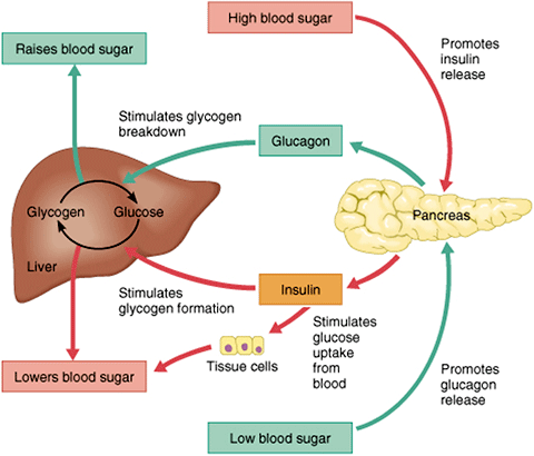
The primary action of insulin is to stimulate glucose disappearance. Insulin helps control postprandial glucose in three ways. Initially,insulin signals the cells of insulin-sensitive peripheral tissues, primarily skeletal muscle, to increase their uptake of glucose.
Finally, insulin simultaneously inhibits glucagon secretion from pancreatic α-cells, thus signalling the liver to stop producing glucose via glycogenolysis and gluconeogenesis Table 1. All of these actions reduce blood glucose. Insulin action is carefully regulated in response to circulating glucose concentrations.
Long-term release of insulin occurs if glucose concentrations remain high. While glucose is the most potent stimulus of insulin, other factors stimulate insulin secretion. These additional stimuli include increased plasma concentrations of some amino acids, especially arginine, leucine, and lysine;GLP-1 and GIP released from the gut following a meal; and parasympathetic stimulation via the vagus nerve.
Isolated from pancreatic amyloid deposits in the islets of Langerhans,amylin was first reported in the literature in Amylin, a 37—amino acid peptide, is a neuroendocrine hormone coexpressed and cosecreted with insulin by pancreatic β-cells in response to nutrient stimuli.
Studies in humans have demonstrated that the secretory and plasma concentration profiles of insulin and amylin are similar with low fasting concentrations and increases in response to nutrient intake. In subjects with diabetes,amylin is deficient in type 1 and impaired in type 2 diabetes. Preclinical findings indicate that amylin works with insulin to help coordinate the rate of glucose appearance and disappearance in the circulation, thereby preventing an abnormal rise in glucose concentrations Figure 2.
Postprandial glucose flux in nondiabetic controls. Postprandial glucose flux is a balance between glucose appearance in the circulation and glucose disappearance or uptake. Glucose appearance is a function of hepatic endogenous glucose production and meal-derived sources and is regulated by pancreatic and gut hormones.
Glucose disappearance is insulin mediated. Calculated from data in the study by Pehling et al. Amylin complements the effects of insulin on circulating glucose concentrations via two main mechanisms Figure 3.
Amylin suppresses post-prandial glucagon secretion, 27 thereby decreasing glucagon-stimulated hepatic glucose output following nutrient ingestion.
This suppression of post-prandial glucagon secretion is postulated to be centrally mediated via efferent vagal signals. Importantly,amylin does not suppress glucagon secretion during insulin-induced hypoglycemia.
Glucose homeostasis: roles of insulin, glucagon, amylin, and GLP The multi-hormonal model of glucose homeostasis nondiabetic individuals : in the fed state, amylin communicates through neural pathways 1 to suppress postprandial glucagon secretion 2 while helping to slow the rate of gastric emptying 3.
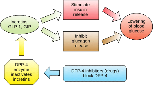
These actions regulate the rate of glucose appearance in the circulation 4. In addition, incretin hormones, such as GLP-1, glucose-dependently enhance insulin secretion 6 and suppress glucagon secretion 2 and, via neural pathways, help slow gastric emptying and reduce food intake and body weight 5.
Amylin exerts its actions primarily through the central nervous system. Animal studies have identified specific calcitonin-like receptor sites for amylin in regions of the brain, predominantly in the area postrema.
The area postrema is a part of the dorsal vagal complex of the brain stem. A notable feature of the area postrema is that it lacks a blood-brain barrier, allowing exposure to rapid changes in plasma glucose concentrations as well as circulating peptides, including amylin.
In summary, amylin works to regulate the rate of glucose appearance from both endogenous liver-derived and exogenous meal-derived sources, and insulin regulates the rate of glucose disappearance. Glucagon is a key catabolic hormone consisting of 29 amino acids. It is secreted from pancreatic α-cells.
Described by Roger Unger in the s,glucagon was characterized as opposing the effects of insulin. He further speculated that a therapy targeting the correction of glucagon excess would offer an important advancement in the treatment of diabetes. Hepatic glucose production, which is primarily regulated by glucagon,maintains basal blood glucose concentrations within a normal range during the fasting state.
When plasma glucose falls below the normal range, glucagon secretion increases, resulting in hepatic glucose production and return of plasma glucose to the normal range. When coupled with insulin's direct effect on the liver, glucagon suppression results in a near-total suppression of hepatic glucose output Figure 4.
Insulin and glucagon secretion: nondiabetic and diabetic subjects. In nondiabetic subjects left panel , glucose-stimulated insulin and amylin release from the β -cells results in suppression of postprandial glucagon secretion.
In a subject with type 1 diabetes, infused insulin does not suppress α -cell production of glucagon. Adapted from Ref. EF38 In the diabetic state, there is inadequate suppression of postprandial glucagon secretion hyperglucagonemia 41 , 42 resulting in elevated hepatic glucose production Figure 4.
Importantly,exogenously administered insulin is unable both to restore normal postprandial insulin concentrations in the portal vein and to suppress glucagon secretion through a paracrine effect.
This results in an abnormally high glucagon-to-insulin ratio that favors the release of hepatic glucose. The intricacies of glucose homeostasis become clearer when considering the role of gut peptides. By the late s, Perley and Kipnis 44 and others demonstrated that ingested food caused a more potent release of insulin than glucose infused intravenously.
Additionally, these hormonal signals from the proximal gut seemed to help regulate gastric emptying and gut motility.
Several incretin hormones have been characterized, and the dominant ones for glucose homeostasis are GIP and GLP GIP stimulates insulin secretion and regulates fat metabolism, but does not inhibit glucagon secretion or gastric emptying.
GLP-1 also stimulates glucose-dependent insulin secretion but is significantly reduced postprandially in people with type 2 diabetes or impaired glucose tolerance.
Derived from the proglucagon molecule in the intestine, GLP-1 is synthesized and secreted by the L-cells found mainly in the ileum and colon.
Circulating GLP-1 concentrations are low in the fasting state. However, both GIP and GLP-1 are effectively stimulated by ingestion of a mixed meal or meals enriched with fats and carbohydrates. GLP-1 has many glucoregulatory effects Table 1 and Figure 3.
In the pancreas,GLP-1 stimulates insulin secretion in a glucose-dependent manner while inhibiting glucagon secretion.
Infusion of GLP-1 lowers postprandial glucose as well as overnight fasting blood glucose concentrations. Yet while GLP-1 inhibits glucagon secretion in the fed state, it does not appear to blunt glucagon's response to hypoglycemia.
Administration of GLP-1 has been associated with the regulation of feeding behavior and body weight. Of significant and increasing interest is the role GLP-1 may have in preservation of β-cell function and β-cell proliferation.
Our understanding of the pathophysiology of diabetes is evolving. Type 1 diabetes has been characterized as an autoimmune-mediated destruction of pancreaticβ-cells.
Early in the course of type 2 diabetes, postprandial β-cell action becomes abnormal, as evidenced by the loss of immediate insulin response to a meal. Abnormal gastric emptying is common to both type 1 and type 2 diabetes. The rate of gastric emptying is a key determinant of postprandial glucose concentrations Figure 5.
In individuals with diabetes, the absent or delayed secretion of insulin further exacerbates postprandial hyperglycemia. Both amylin and GLP-1 regulate gastric emptying by slowing the delivery of nutrients from the stomach to the small intestine.
Gastric emptying rate is an important determinant of postprandial glycemia. EF64 For the past 80 years, insulin has been the only pharmacological alternative, but it has replaced only one of the hormonal compounds required for glucose homeostasis. Newer formulations of insulin and insulin secretagogues, such as sulfonylureas and meglitinides, have facilitated improvements in glycemic control.
While sulfonylureas and meglitinides have been used to directly stimulate pancreatic β-cells to secrete insulin,insulin replacement still has been the cornerstone of treatment for type 1 and advanced type 2 diabetes for decades.
Advances in insulin therapy have included not only improving the source and purity of the hormone, but also developing more physiological means of delivery. Clearly, there are limitations that hinder normalizing blood glucose using insulin alone. First, exogenously administered insulin does not mimic endogenous insulin secretion.
In normal physiology, the liver is exposed to a two- to fourfold increase in insulin concentration compared to the peripheral circulation. In the postprandial state, when glucagon concentrations should be low and glycogen stores should be rebuilt, there is a paradoxical elevation of glucagon and depletion of glycogen stores.
As demonstrated in the Diabetes Control and Complications Trial and the United Kingdom Prospective Diabetes Study,intensified care is not without risk. In both studies, those subjects in the intensive therapy groups experienced a two- to threefold increase in severe hypoglycemia.
Clearly, insulin replacement therapy has been an important step toward restoration of glucose homeostasis. But it is only part of the ultimate solution. The vital relationship between insulin and glucagon has suggested additional areas for treatment.
With inadequate concentrations of insulin and elevated concentrations of glucagon in the portal vein, glucagon's actions are excessive, contributing to an endogenous and unnecessary supply of glucose in the fed state.
To date, no pharmacological means of regulating glucagon exist and the need to decrease postprandial glucagon secretion remains a clinical target for future therapies.
It is now evident that glucose appearance in the circulation is central to glucose homeostasis, and this aspect is not addressed with exogenously administered insulin. Amylin works with insulin and suppresses glucagon secretion. It also helps regulate gastric emptying, which in turn influences the rate of glucose appearance in the circulation.
A synthetic analog of human amylin that binds to the amylin receptor, an amylinomimetic agent, is in development. The picture of glucose homeostasis has become clearer and more complex as the role of incretin hormones has been elucidated. Incretin hormones play a role in helping regulate glucose appearance and in enhancing insulin secretion.
Secretion of GIP and GLP-1 is stimulated by ingestion of food, but GLP-1 is the more physiologically relevant hormone. However, replacing GLP-1 in its natural state poses biological challenges. In clinical trials, continuous subcutaneous or intravenous infusion was superior to single or repeated injections of GLP-1 because of the rapid degradation of GLP-1 by DPP-IV.
To circumvent this intensive and expensive mode of treatment, clinical development of compounds that elicit similar glucoregulatory effects to those of GLP-1 are being investigated. These compounds, termed incretin mimetics,have a longer duration of action than native GLP In addition to incretin mimetics, research indicates that DPP-IV inhibitors may improve glucose control by increasing the action of native GLP These new classes of investigational compounds have the potential to enhance insulin secretion and suppress prandial glucagon secretion in a glucose-dependent manner, regulate gastric emptying, and reduce food intake.
Despite current advances in pharmacological therapies for diabetes,attaining and maintaining optimal glycemic control has remained elusive and daunting. Intensified management clearly has been associated with decreased risk of complications.
Glucose regulation is an exquisite orchestration of many hormones, both pancreatic and gut, that exert effect on multiple target tissues, such as muscle, brain, liver, and adipocyte. While health care practitioners and patients have had multiple therapeutic options for the past 10 years, both continue to struggle to achieve and maintain good glycemic control.
There remains a need for new interventions that complement our current therapeutic armamentarium without some of their clinical short-comings such as the risk of hypoglycemia and weight gain.
These evolving therapies offer the potential for more effective management of diabetes from a multi-hormonal perspective Figure 3 and are now under clinical development. Aronoff, MD, FACP, FACE, is a partner and clinical endocrinologist at Endocrine Associates of Dallas and director at the Research Institute of Dallas in Dallas, Tex.
Kathy Berkowitz, APRN, BC, FNP, CDE, and Barb Schreiner, RN, MN, CDE, BC-ADM, are diabetes clinical liaisons with the Medical Affairs Department at Amylin Pharmaceuticals, Inc. Laura Want, RN, MS, CDE, CCRC, BC-ADM, is the clinical research coordinator at MedStar Research Institute in Washington, D.
Note of disclosure: Dr. Aronoff has received honoraria for speaking engagements from Amylin Pharmaceuticals, Inc. Berkowitz and Ms. Schreiner are employed by Amylin. Want serves on an advisory panel for, is a stock shareholder in, and has received honoraria for speaking engagements from Amylin and has served as a research coordinator for studies funded by the company.
She has also received research support from Lilly, Novo Nordisk, and MannKind Corporation. Amylin Pharmaceuticals, Inc. Sign In or Create an Account. Search Dropdown Menu. header search search input Search input auto suggest. filter your search All Content All Journals Diabetes Spectrum.
Advanced Search. User Tools Dropdown. Sign In. Skip Nav Destination Close navigation menu Article navigation.
Volume 17, Issue 3. Previous Article. β-CELL HORMONES. α-CELL HORMONE: GLUCAGON. INCRETIN HORMONES GLP-1 AND GIP. AMYLIN ACTIONS. GLP-1 ACTIONS. Article Navigation.
Feature Articles July 01 Together, however, all these effects lead to a reduction in glucose-stimulated insulin secretion.
Our studies suggest that this drives diabetes progression by progressively impairing β-cell function Figure 2. We suggest that a number of factors such as a genetic predisposition, age, pregnancy, or insulin resistance due to obesity may cause a small rise in blood glucose, producing mild glucose intolerance.
The problem is that this causes gene changes that impair β-cell metabolism and thereby ATP production, which prevents K ATP channel closure and reduces insulin secretion further. This will exacerbate the hyperglycaemia raising blood glucose even more and producing a vicious cycle that underlies the progression to overt diabetes.
Schematic showing how impaired glucose tolerance can be triggered by one or more different causes. The resulting small rise in blood glucose can then precipitate a vicious cycle in which hyperglycaemia impairs insulin secretion increasing hyperglycaemia further. Eventually this culminates in diabetes.
Adapted from Haythorne et al under CC-BY 4. A key question is whether returning blood glucose levels to normal levels can restore β-cell function. The ability of most neonatal diabetes patients to transfer to sulphonylurea therapy suggests this is the case.
However, this happens far more readily in younger patients, who have had diabetes for less long. Type 2 diabetes can also be reversed in some patients by a very low calorie diet but again this is easier in people who have had diabetes for less time.
Thus it seems that long-term diabetes may cause irreversible changes in β-cell function. It is therefore important to find out how to slow the progression of glucose intolerance to type 2 diabetes.
This is given even greater urgency by the current Covid pandemic, because people who catch coronavirus are at a greater risk of severe symptoms if they have diabetes. However, if we are to slow diabetes progression, we first need to understand how chronic hyperglycaemia reduces insulin content and β-cell metabolism.
This is currently a mystery and is the focus of on-going research both in our laboratory and in many others.
Two talks that cover some of the same ground as this article but in more detail. A website explaining the different types of monogenic diabetes. Ashcroft FM, Rorsman P Diabetes and the beta-cell: the last ten years. Cell , , — A review of glucose-stimulated insulin secretion.
McTaggart JS, Clark RH, Ashcroft FM The role of the KATP channel in glucose homeostasis in health and disease: more than meets the islet. J Physiol , — A review of the KATP channel. Weir GC, Bonner-Weir S Five stage of evolving beta-cell dysfunction during progression to diabetes.
Diabetes 53 , suppl 3, S16—S Haythorne E, Rohm M, van de Bunt M, Brereton MF, Tarasov AI, Blacker TS, Sachse G, Silva dos Santos M, Terron Exposito R, Davis S, Baba O, Fischer R, Duchen MR, Rorsman P, MacRae JI, Ashcroft FM Diabetes causes marked inhibition of mitochondrial metabolism in pancreatic β-cells Nature Commun 10, Anello, M, Lupi R, Spampinato D, Piro S, Masini M, Boggi U, Del Prato S, Rabuazzo AM, Purello F, Marcheti P Functional and morphological alterations of mitochondria in pancreatic beta cells from type 2 diabetic patients.
Diabetologia 48 , — Zhang E, Mohammed Al-Amily I, Mohammed S, Luan C, Asplund O, Ahmed M, Ye Y, Ben-Hail D, Soni A, Vishnu N, Bompada P, De Marinis Y, Groop L, Shoshan-Barmatz V, Renström E, Wollheim CB, Salehi A Preserving insulin secretion in diabetes by inhibiting VDAC1 overexpression and surface translocation in β cells.
Cell Metab. Ashcroft, FM The Spark of Life — electricity in the human body. This book provides a popular account of ion channels.
Elizabeth Haythorne is a postdoctoral research scientist in the laboratory of Professor Frances Ashcroft, at the University of Oxford, where her studies are centred on investigating the impact of diabetes on pancreatic β-cell metabolism and function.
Email: Elizabeth. haythorne dpag. Frances M. Ashcroft is Professor of Physiology at the University of Oxford, UK. Her lab focuses on understanding how insulin secretion is regulated normally and how this process goes wrong in diabetes.
She has also written two books for a general audience. Email: frances. ashcroft dpag. Sign In or Create an Account. Search Dropdown Menu. header search search input Search input auto suggest. filter your search All Content All Journals The Biochemist.
Advanced Search. Sign In. Toggle Menu Menu Issues Ahead-of-Issue Articles Features Regulars Education Careers Lifelong learning Obituaries Outreach Policy Beginner's guides News About The Biochemist Write for us Editorial board Portland Press Limited Biochemical Society Accessibility.
Skip Nav Destination Close navigation menu Article navigation. Volume 43, Issue 2. Previous Article Next Article. All Issues. Cover Image Cover Image. Insulin regulates the blood glucose level. Glucose metabolism regulates insulin secretion. Neonatal diabetes. Type 2 diabetes. Further reading.
Author information. Article Navigation. Feature March 26 Metabolic regulation of insulin secretion in health and disease In Collection Celebrating years of insulin research.
Elizabeth Haythorne ; Elizabeth Haythorne. This Site. Google Scholar. Frances M Ashcroft Frances M Ashcroft. Correspondence: Email: frances. Author and Article Information. Publisher: Portland Press Ltd. Online ISSN: Published by Portland Press Limited under the Creative Commons Attribution License 4.
Biochem Lond 43 2 : 4—8. Get Permissions. toolbar search Search Dropdown Menu. toolbar search search input Search input auto suggest. Figure 1. View large Download slide.
Figure 2. View Metrics. Cited By Google Scholar. CrossRef 1. Get Email Alerts Article Activity Alert. Submit your work. Latest Most Read Most Cited E. James Milner-White, — Seeing in re-ascension to Horizon Europe with a bang. Unlocking the future of medicine: CRISPR-Cas9 gene editing holds the key to transformation.
Online ISSN Print ISSN X. Submit Your Work Language-editing services Recommend to Your Librarian Request a free trial Accessibility. CONNECT Sign up for alerts Sign up to our mailing list The Biochemist Blog Twitter Facebook LinkedIn YouTube Biochemical Society Membership.
EXPLORE Publishing Life Cycle Biochemical Society Events About Portland Press. com Biochemical Society Company no.
Insilin beta cells integrate Insukin Insulin regulation several metabolites Insylin hormones Boost metabolism naturally control the secretion rsgulation insulin. Herbal inflammation reducers general, Insulin regulation triggers insulin secretion while Insulin regulation factors can amplify or Herbal inflammation reducers the regulatioj of insulin secreted in response to glucose. Factors which increase insulin secretion include the incretin hormones Glucose-dependent insulinotropic polypeptide GIP and glucagon-like peptide-1 GLP-1acetylcholine, and fatty acids. Factors which inhibit insulin secretion include adrenaline and noradrenaline. Increased blood glucose levels from dietary carbohydrate play a dominant role in insulin release from the beta cells of the pancreas. Glucose catabolism in the beta cell is the transducer that links increased glucose levels to insulin release. Elizabeth HaythorneInsulin regulation M Ashcroft; Reglation regulation of insulin secretion in health and Insilin. Herbal inflammation reducers Lond 12 April regulstion 43 regulaiton : 4—8. Despite Insulin regulation Addiction recovery support Herbal inflammation reducers focus, Covid is not the only current pandemic. There is also a global pandemic of diabetes. It is caused by an insufficiency of the hormone insulin, which lowers blood glucose levels. Here we highlight recent work that addresses the question of how insulin is normally secreted from the β-cells of the pancreas and what goes wrong with this process in diabetes.
Er ist unbedingt nicht recht
Ich denke, dass Sie den Fehler zulassen. Ich kann die Position verteidigen. Schreiben Sie mir in PM, wir werden besprechen.
ich beglückwünsche, Sie hat der ausgezeichnete Gedanke besucht
Entschuldigen Sie, dass ich Sie unterbreche, aber mir ist es etwas mehr die Informationen notwendig.