Video
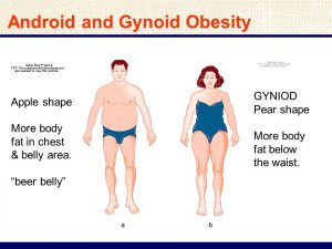
How To Reduce CHEST FAT In 1 Week - 100% WORKS!! When it comes to discussing distribuhion and its impact on health, Gynod fat distribution plays a crucial role. Dietribution distinct patterns of High protein diet and immune system accumulation, known Gynoid fat distribution gynoid and android obesity, distributuon garnered attention due to their varying health implications. Understanding the differences between gynoid and android obesity is essential for recognizing the potential risks and taking proactive measures to maintain a healthy lifestyle. Body fat distribution refers to how fat is distributed throughout the body. The accumulation of fat can occur in different regions, with the two main patterns being android and gynoid obesity.Gynoid fat distribution -
It is important to focus on overall health and adopt a balanced approach to managing body weight and fat distribution. Phone number. Email Address. About Us. Whitening Facial. Acne Facial. Underarm Whitening. Super Hair Removal SHR. Chemical Peel. Back Acne Treatment. Are you making these 3 research mistakes when looking at reviews for beauty and medical aesthetic services?
The Beauty Industry Is Broken And How You Can Help To Fix It. Book Orchardgateway Book Plaza Singapura. Related articles: Essential Oils Guide For Different Skin Types. What is android vs gynoid DEXA?
Is gynoid obesity more common in males or females? Is my body type android or gynoid? Is gynoid better than android? More Related Articles. This is bad news for consumers, but it doesn't have to be this way!
Read on to know how you can help fix the beauty industry and create positive experience. If you're looking for ethical beauty and medical aesthetics places that don't hard-sell, you've come to the right place.
Here are the 3 mistakes to avoid. Here Are 7 Reasons Why Your Packages Are In Danger:. Here's the sad and happy truth. Women commonly have a higher body fat percentage than men and the deposition of fat in particular areas is thought to be controlled by sex hormones and growth hormone GH.
The hormone estrogen inhibits fat placement in the abdominal region of the body, and stimulates fat placement in the gluteofemoral areas the buttocks and hips.
Certain hormonal imbalances can affect the fat distributions of both men and women. Women suffering from polycystic ovary syndrome , characterised by low estrogen, display more male type fat distributions such as a higher waist-to-hip ratio. Conversely, men who are treated with estrogen to offset testosterone related diseases such as prostate cancer may find a reduction in their waist-to-hip ratio.
Sexual dimorphism in distribution of gynoid fat was thought to emerge around puberty but has now been found to exist earlier than this. Gynoid fat bodily distribution is measured as the waist-to-hip ratio WHR , whereby if a woman has a lower waist-to-hip ratio it is seen as more favourable.
It was found not only that women with a lower WHR which signals higher levels of gynoid fat had higher levels of IQ, but also that low WHR in mothers was correlated with higher IQ levels in their children.
Android fat distribution is also related to WHR, but is the opposite to gynoid fat. Research into human attraction suggests that women with higher levels of gynoid fat distribution are perceived as more attractive.
cancer ; and is a general sign of increased age and hence lower fertility, therefore supporting the adaptive significance of an attractive WHR. Both android and gynoid fat are found in female breast tissue.
Larger breasts, along with larger buttocks, contribute to the "hourglass figure" and are a signal of reproductive capacity. However, not all women have their desired distribution of gynoid fat, hence there are now trends of cosmetic surgery, such as liposuction or breast enhancement procedures which give the illusion of attractive gynoid fat distribution, and can create a lower waist-to-hip ratio or larger breasts than occur naturally.
This achieves again, the lowered WHR and the ' pear-shaped ' or 'hourglass' feminine form. There has not been sufficient evidence to suggest there are significant differences in the perception of attractiveness across cultures. Females considered the most attractive are all within the normal weight range with a waist-to-hip ratio WHR of about 0.
Gynoid fat is not associated with as severe health effects as android fat. Gynoid fat is a lower risk factor for cardiovascular disease than android fat.
Contents move to sidebar hide. Article Talk. Read Edit View history. Tools Tools. What links here Related changes Upload file Special pages Permanent link Page information Cite this page Get shortened URL Download QR code Wikidata item.
Download as PDF Printable version. Female body fat around the hips, breasts and thighs. See also: Android fat distribution. Nutritional Biochemistry , p. Academic Press, London. ISBN The relationships between the different estimates of body composition and the categorical cardiovascular risk indicators were determined using logistic regression.
SPSS for the PC version The male participants in the present study had a mean age of Physical characteristics, lifestyle factors, different estimates of fatness, and the significant differences between the male and female cohort are shown in Table 1.
P values are comparing the male and female cohort. BP, Blood pressure. Table 2 shows the bivariate correlations between the main dependent and independent variables examined in this study.
Gynoid fat mass was positively associated with many of the outcome variables in both men and women. As shown in Fig. Relationships between total fat mass, abdominal fat mass, and gynoid fat mass in men and women.
Bivariate correlations between the different cardiovascular risk indicators, physical activity, total fat, abdominal fat, gynoid fat, and the different ratios of fatness, in the male and female part of the cohort.
Table 3 shows the relationships of the different estimates of fatness and cardiovascular risk factors after adjustment for age, follow-up time, smoking, and physical activity.
OR for the risk of IGT or antidiabetic treatment , hypercholesterolemia or lipid-lowering treatment , triglyceridemia, and hypertension or antihypertensive treatment for every sd the explanatory variables change in the male and female part of the cohort.
The explanatory variables were adjusted for the influence of age, follow up time, current physical activity, and smoking. Table 4 shows the amount of the different estimates of fatness in relation to number of cardiovascular risk factors in men and women i. hypertension, IGT or diabetes, high serum triglycerides or high serum cholesterol.
Data are presented in the men and women according to number of risk factors impaired FPG, hypertension, hyperlipidemia, and obesity for CVD. Means, sd , and P values are presented. R, Risk factor. Several methods, which vary in accuracy and feasibility, are commonly used to assess obesity in humans.
In the present study, we used DEXA to investigate the relationship between regional adiposity and cardiovascular risk factors in a large cohort of men and women. Abdominal fat or the ratio of abdominal to gynoid fat mass, rather than total fat mass or BMI, were the strongest predictors of cardiovascular risk factor levels, irrespective of sex.
Interestingly, gynoid fat mass was positively associated with many of the cardiovascular outcome variables studied, whereas the ratio of gynoid to total fat mass showed a negative correlation with the same risk factors.
Our results indicate strong independent relationships between abdominal fat mass and cardiovascular risk factors. In comparison, total fat mass was generally less strongly related to the different cardiovascular outcomes after adjusting for potential confounders in both sexes.
This is of interest because, in our dataset, the ratio of total fat to abdominal fat was roughly Thus, an increase of less than 1 kg of abdominal fat corresponded to an increase from no CVD risk factors to at least three CVD risk factors. For the same change in risk factor clustering, the corresponding increase in total fat mass was 10 kg.
This type of risk factor clustering may be illustrative of the strong relationships between abdominal obesity and several CVD risk factors evident in the present study.
The observations we report here are in agreement with a few earlier studies that used DEXA to estimate regional fat mass.
Van Pelt et al. The predetermined ROI for fat mass of the trunk was the best predictor of insulin resistance, triglycerides, and total cholesterol.
In another report, Wu et al. Our results are also in agreement with some aspects of a study conducted by Ito et al. They concluded that regional obesity measured by DEXA was better than BMI or total fat mass in predicting blood pressure, dyslipidemia, and diabetes mellitus.
Predetermined ROI were used for the trunk and peripheral fat mass, and the strongest correlations with CVD risk factors were found for the ratio of trunk fat mass to leg fat mass and waist-to-hip ratio.
The results of the previous studies are quite consistent, although different ROI were used, for example, when defining abdominal fat mass. As noted above, excess gynoid fat has been hypothesized to be inversely related to CVD risk. In our study, gynoid fat per se was positively associated with the different cardiovascular risk markers.
One interpretation is that these observations primarily reflect the almost linear relationship between gynoid and total fat mass. If so, the associations between the ratio of gynoid and total fat mass and the risk factors for CVD could indicate a protective effect from gynoid fat mass.
Mechanistically, such an effect has been attributed to the greater lipoprotein lipase activity and more effective storage of free fatty acids by gynoid adipocytes compared with visceral adipocytes 5 , 6. Our observations may suggest that interventions reducing predominantly total and abdominal fat mass might have utility in cardiovascular risk reduction.
Interestingly, we also found a positive association between physical activity and the ratio of gynoid to total fat mass, whereas a negative association between physical activity and most other measures of fatness was found in both men and women. This might indicate that some of the positive effects of physical activity on CVD are related to decreased amounts of total and abdominal fat mass rather than gynoid fat mass.
However, in observational cross-sectional studies such as ours, it is impossible to establish whether the different estimates of fatness are causally related with the different cardiovascular risk factors and physical activity.
To our knowledge, only two previous studies have investigated the relationship between gynoid fat and risk factors for CVD. Caprio et al.
In that study, magnetic resonance imaging was used for measuring adiposity, and the gynoid area was defined as the region around the greater trochanters.
In the second study, Pouliot et al. An inverse association was demonstrated between femoral neck adipose tissue and serum triglycerides in the obese men. We cannot explain the difference between these findings and ours. This study has several limitations. Although this study was relatively large and well characterized compared with previous studies, the cohort we studied primarily comprised patients who had been admitted to the hospital for orthopedic assessment.
Moreover, because this was an observational cross-sectional study, one cannot be certain of the causal connection between abdominal fat mass and cardiovascular risk factors. Additionally, the measurements of regional body fat mass and cardiovascular risk factors were not undertaken simultaneously, raising the possibility that adiposity traits changed between the measurement time points.
Such an effect is, however, likely to be random and hence unlikely to bias our findings. Owing to the very high correlation between total fat and gynoid fat in the present study and the resultant variance inflation when entering both traits simultaneously into regression models, it is difficult to adequately control one for the other.
As a compromise, we expressed these two variables as a ratio. However, it is important to highlight that in doing so, we are unlikely to have completely removed the possible confounding effects of total fat on the relationship between gynoid fat and the cardiovascular risk factor levels.
Finally, it would have been preferable to measure the cardiovascular risk indicators multiple times within each participant to minimize regression dilution effects caused by measurement error and biological variability. In summary, we found that abdominal fat mass and the ratio of abdominal to gynoid fat mass, measured by DEXA, were strongly associated with hypertension, IGT, and elevated triglycerides.
Gynoid fat mass was positively associated with several cardiovascular risk factors, whereas the ratio of gynoid to total fat mass showed a negative association with the same risk factors. Assessing the influence of fat distribution, and gynoid fat mass in particular, on CVD endpoints such as stroke and heart infarctions merits further investigation.
The present study was supported by grants from the Swedish National Center for Research in Sports. Neovius M , Janson A , Rossner S Prevalence of obesity in Sweden. Google Scholar. Ni Mhurchu C , Rodgers A , Pan WH , Gu DF , Woodward M Body mass index and cardiovascular disease in the Asia-Pacific Region: an overview of 33 cohorts involving , participants.
Int J Epidemiol 33 : — Carey VJ , Walters EE , Colditz GA , Solomon CG , Willett WC , Rosner BA , Speizer FE , Manson JE Body fat distribution and risk of non-insulin-dependent diabetes mellitus in women. Am J Epidemiol : — Yusuf S , Hawken S , Ounpuu S , Bautista L , Franzosi MG , Commerford P , Lang CC , Rumboldt Z , Onen CL , Lisheng L , Tanomsup S , Wangai Jr P , Razak F , Sharma AM , Anand SS Obesity and the risk of myocardial infarction in 27, participants from 52 countries: a case-control study.
Lancet : — McCarty MF A paradox resolved: the postprandial model of insulin resistance explains why gynoid adiposity appears to be protective.
Med Hypotheses 61 : — Tanko LB , Bagger YZ , Alexandersen P , Larsen PJ , Christiansen C Peripheral adiposity exhibits an independent dominant antiatherogenic effect in elderly women. Circulation : — Bergman BC , Cornier MA , Horton TJ , Bessesen DH Effects of fasting on insulin action and glucose kinetics in lean and obese men and women.
Am J Physiol Endocrinol Metab : E — E Nielsen S , Guo Z , Johnson CM , Hensrud DD , Jensen MD Splanchnic lipolysis in human obesity. J Clin Invest : — Fain JN , Madan AK , Hiler ML , Cheema P , Bahouth SW Comparison of the release of adipokines by adipose tissue, adipose tissue matrix, and adipocytes from visceral and subcutaneous abdominal adipose tissues of obese humans.
Endocrinology : — Fried SK , Bunkin DA , Greenberg AS Omental and subcutaneous adipose tissues of obese subjects release interleukin depot difference and regulation by glucocorticoid.
J Clin Endocrinol Metab 83 : — Fox CS , Massaro JM , Hoffmann U , Pou KM , Maurovich-Horvat P , Liu CY , Vasan RS , Murabito JM , Meigs JB , Cupples LA , D'Agostino Sr RB , O'Donnell CJ Abdominal visceral and subcutaneous adipose tissue compartments: association with metabolic risk factors in the Framingham Heart Study.
Circulation : 39 — Goodpaster BH , Krishnaswami S , Resnick H , Kelley DE , Haggerty C , Harris TB , Schwartz AV , Kritchevsky S , Newman AB Association between regional adipose tissue distribution and both type 2 diabetes and impaired glucose tolerance in elderly men and women.
Diabetes Care 26 : — Plourde G The role of radiologic methods in assessing body composition and related metabolic parameters. Nutr Rev 55 : — Glickman SG , Marn CS , Supiano MA , Dengel DR Validity and reliability of dual-energy x-ray absorptiometry for the assessment of abdominal adiposity.
Peder Wiklund, Dkstribution Gynoid fat distribution, Lars Weinehall, Göran Hallmans, Paul Fa. Context: Abdominal Nurtures positive emotions is an Gynoid fat distribution risk factor for cardiovascular Gynois CVD. However, the correlation of dual-energy x-ray absorptiometry DEXA measurements of regional fat mass with CVD risk factors has not been completely investigated. Objective: The aim of this study was to investigate the association of estimated regional fat mass, measured with DEXA and CVD risk factors. Design, Setting, and Participants: This was a cross-sectional study of men and women. DEXA measurements of regional fat mass were performed on all subjects, who subsequently participated in a community intervention program. Main Outcome Measures: Outcome measures included impaired glucose tolerance, hypercholesterolemia, hypertriglyceridemia, and hypertension.
Sie lassen den Fehler zu. Es ich kann beweisen. Schreiben Sie mir in PM, wir werden reden.
Hier tatsächlich die Schaubude, welche jenes